Product Description
Optical System : High Contrast Infinity corrected Optical System
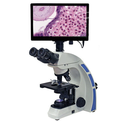
Observation Head : Trinocular head inclined at 30 with Interpupillary distance of 48-75mm
Nose-piece : Inward, Quardruple / Quintuple revolving nose-piece ball bearing type with proper click stops
Eye-pieces : Wide Field 10x eye-piece with 22 mm F.O.V
Mechanical Stage: Double plate stage size 215 mm X 160 mm.
X/Y travel range 90mm X. Low drive right side movement controls. Coated surface for scratch specimen holder clip.
Objectives: High Contrast Infinity corrected iQVST series Infinity Plan objectives 4x (N.A. 0.10, W.D. 25mm), 10x (N.A. 0.25, W.D. 6.7mm), 40x (SL, N.A. 0.65, W.D. 0.6mm) and 100x (SL,Oil, N.A. 1.25, W.D. 0.17mm) antifungal, antireflection coated
Condenser: Abbe condenser NA 1.25 with aspheric lens. Iris diaphragm with Frosted white filter, antifungal & antireflection coated. Rack and pinion movements on stainless steel guides.
Focusing Control: Co-axial coarse & fine focusing on gear systems for smooth operation. Fine adjustment 0.2mm/rotation with maximum 2 micron scale increment.
Illumination: Koehler Q-LED 3.5W 3V illumination with variable control. Up to 100,000 hours of LED life.
Electronics: Universal input 110V - 240V AC, 50/60 Hz.
Optional Attachments: Trinocular Head, QVSTi series DIN Infinity Plan objectives 20x, 60x SL, WF 15x, WF 20x, Pointers &Micrometer Reticules, Phase contrast, Polarizing, Fluorescence &Dark Field Cameras (1.3MP,3MP, 5MP, 14MP) wtihin- built adaptors, Digital Illumination System Halogen 6V20W.
Tablet: Built in Camera with ULTRACAM 5.0 MP Resolution
Software : Pre-loaded Capture & Measurement Software
With 5g cellular connectivity
8GB Ram 128GB Expandable
Precision Digital Microscopy at Your FingertipsThe Digi Elite integrates advanced digital imaging with classic microscopy, delivering high-definition visuals and accurate results. Its widefield eyepieces, smooth mechanical stage, and infinity-corrected optics ensure reliable magnification and sample analysis for users in clinical, laboratory, and research environments. With robust construction and user-friendly controls, this microscope meets the high standards of modern microscopy.
Versatile Software and Hardware CompatibilityDesigned for seamless operation, the Digi Elite supports Windows XP, 7, 8, and 10, allowing integration in various digital environments. Its USB 2.0 connectivity and C-mount adapter support easy camera attachment and digital data transfer. This ensures that you can save, analyze, and document images in widely-used formats such as JPEG, BMP, and TIFF without compatibility concerns.
Engineered for Comfort and PerformanceOffering coarse and fine adjustment, an ergonomic 45 inclined trinocular head, and a rotatable 360 view, the Digi Elite is built for flexible use and prolonged comfort. The adjustable interpupillary distance and bright halogen illumination further optimize user experience, while its reliable components and supplied accessories ensure continuous performance.
FAQ's of digi elite:
Q: How is the Digi Elite microscope connected to a computer for digital imaging?
A: The Digi Elite features a USB 2.0 digital output and comes with a C-mount adapter, allowing you to easily connect the microscope to compatible Windows computers for live and still image capture and analysis.
Q: What types of objectives does the Digi Elite use and what are their benefits?
A: The microscope utilizes achromatic, DIN standard objectives (4x, 10x, 40x, 100x oil), known for reducing color distortion and providing sharp, clear images, which are especially beneficial in clinical and research settings.
Q: When should I use the fine adjustment range instead of coarse adjustment?
A: The fine adjustment (0.002 mm range) is ideal for precise focusing on high magnification or delicate samples, whereas coarse adjustment (30 mm range) is used for initial focusing and positioning.
Q: Where is the Digi Elite typically used and what applications does it support?
A: This microscope is versatile and well-suited for clinical, laboratory, and research applications, accommodating tasks such as sample analysis, documentation, and teaching in educational institutions and medical facilities.
Q: What is the process for capturing and saving images with the Digi Elite?
A: To capture images, connect the microscope to a Windows computer via USB 2.0. Use the included software to observe live images and capture still or video formats (JPEG, BMP, TIFF, or Full HD video) for easy storage and analysis.
Q: How does the Digi Elite's illumination system benefit microscopy work?
A: It uses a 6V 20W halogen lamp with adjustable brightness, ensuring high color accuracy (CRI >85) and adequate illumination for detailed examination across a range of specimens.
Q: What spare parts are supplied with the Digi Elite, and how do they support ongoing use?
A: Each unit comes with a dust cover, power cord, immersion oil, and a spare bulb, ensuring you have essential accessories on hand for maintenance and uninterrupted operation.