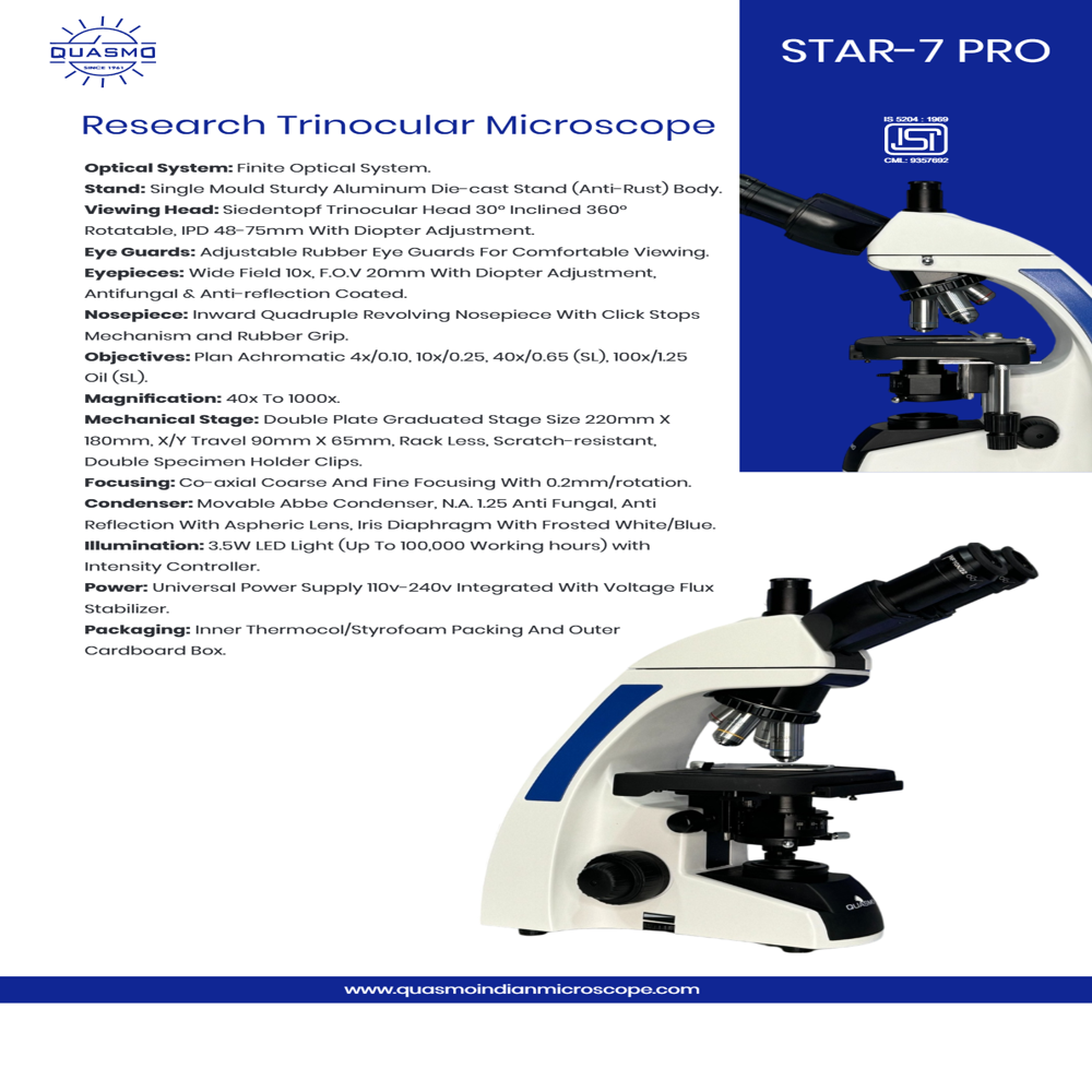
Product Description
SPECIFICATIONS:
Optical System: High Contrast finite colour corrected Optical system
Magnification: 40x to 1000x visiual magnicifation
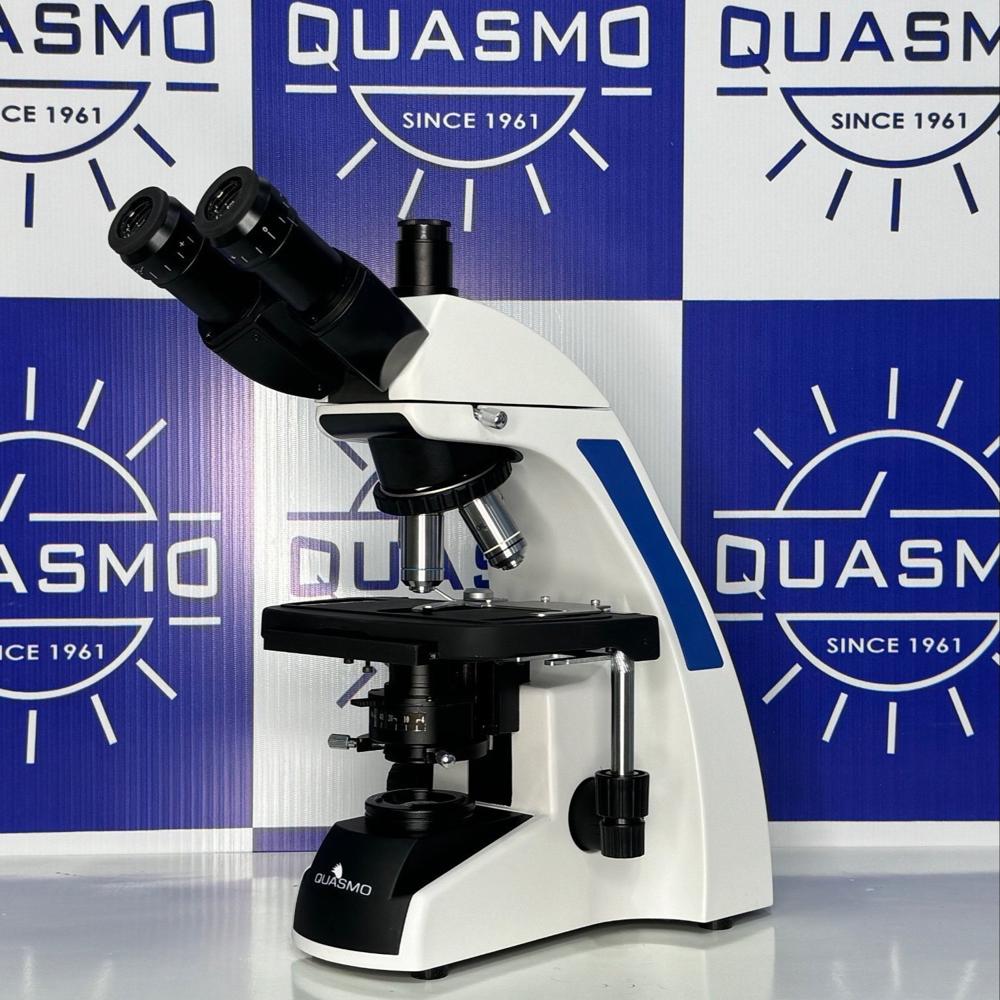
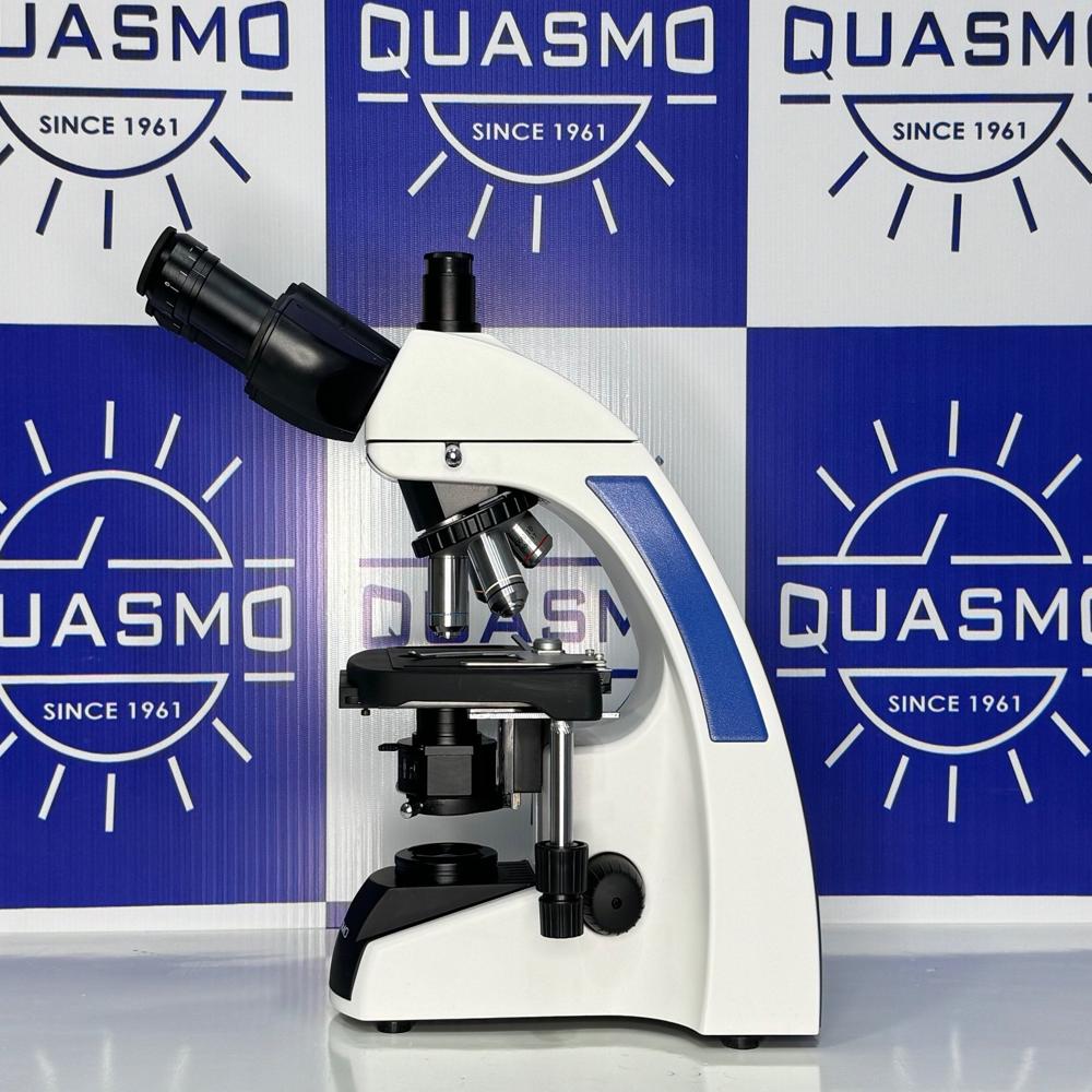
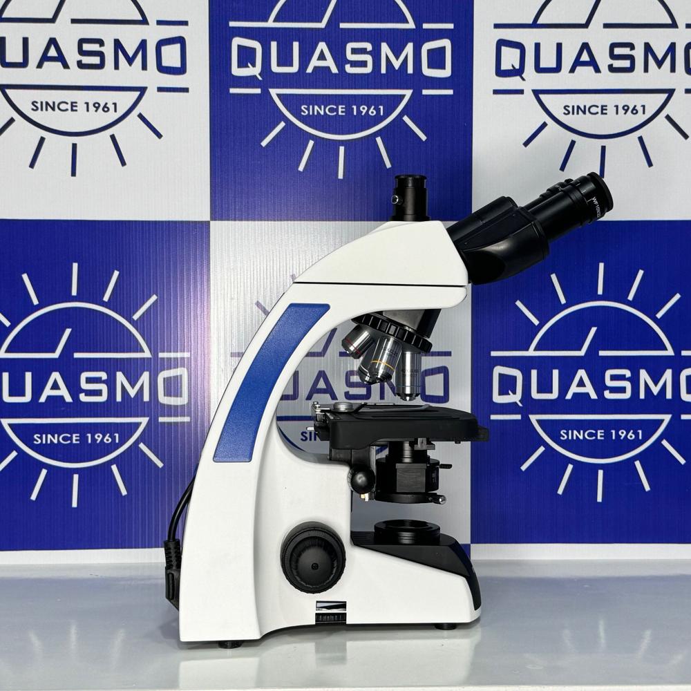
Stand: Aluminium single die-cast stand for enhanced comfort and stability
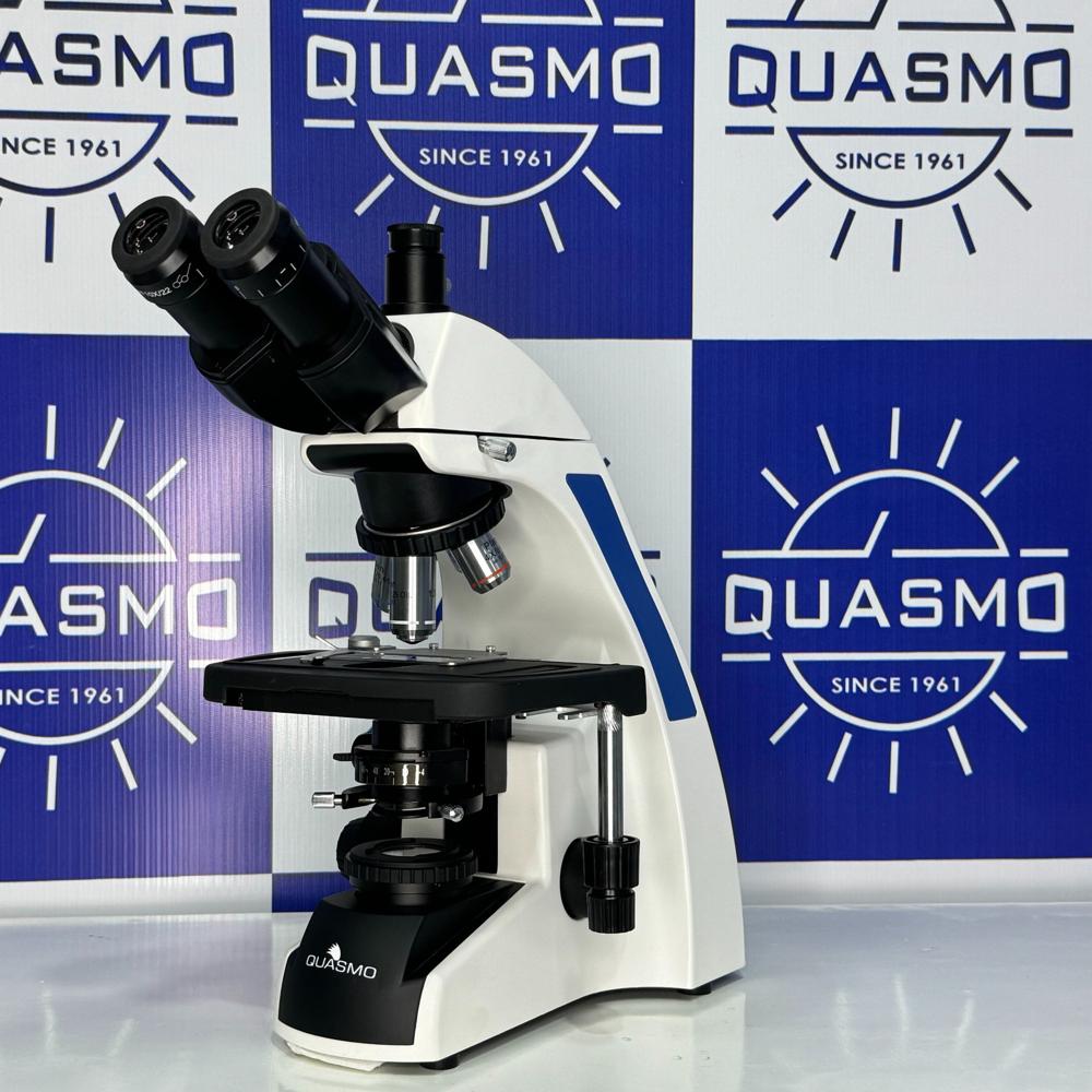
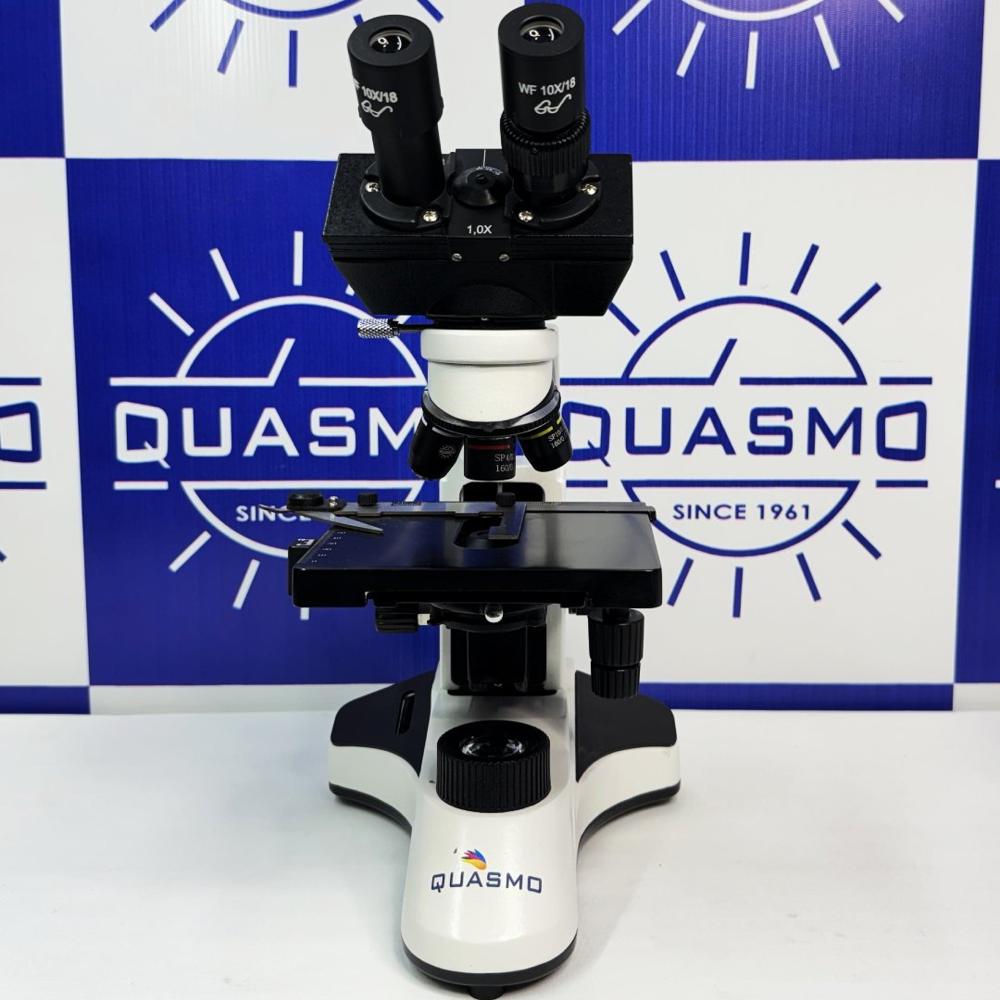
Observation Head: Sidentopf Trinocular head, 30 degree inclined , 360 degree rotatable , interpupilary distance 48-75mm
Eye-pieces: Widefield 10x/20mm with foldable rubber eye guards, antifungal & anti-reflection hard coated with dual diopteric adjustment
Nose-piece: Reverse angle quadruple nose-piece (Ball bearing type) with precise click stops
Objectives: QUSP series high contrast flat field DIN Achromatic objectives4x,10x, 40x(spring loaded) , 100x(spring loaded,oil) , antifungal & anti -reflection hard coated
Mechanical Stage: Double plate stage 220mm X 160mm , X/Y travel range 90mm X 65mm. Low drive right hand movement controls hard coated surface for scratch resistance. Double specimen holder clip.
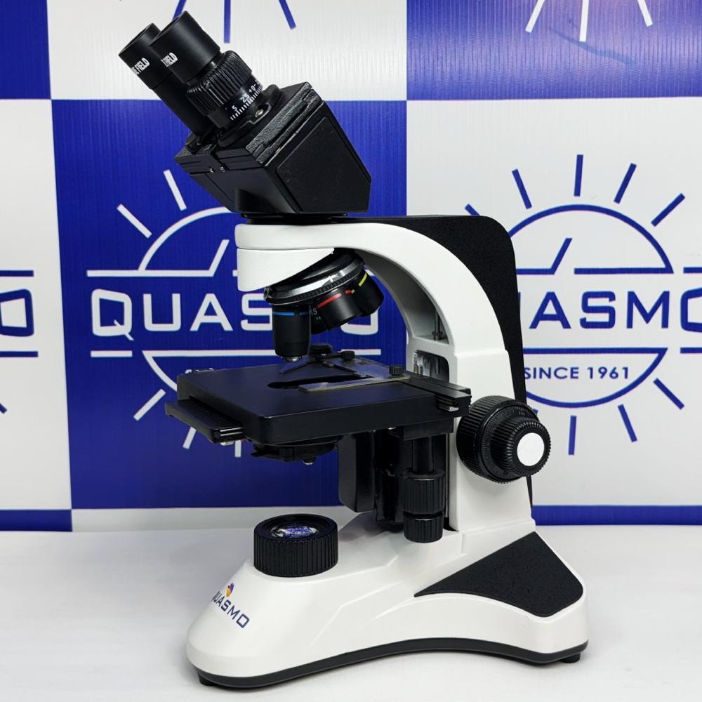
Condenser: Sub-stage Abbe condenser NA 1.25 with aspheric lens. Iris diaphragm with frosted white/blue filter, anti-fungal & anti- reflection coated. Rack and pinion movements on stainless steels guides
Focusing Control: Co-axial coarse and fine focusing on gear systems for smooth operation. Fine adjustment 0.2mm/rotation with maximum 2 micron scale increment.
Illumination: LED 3W illumination with variable illumination control. Up to 100,000 hours of LED Life
Electronics: Universal input 110v-240v AC, 50/60Hz. Built in voltage stabilizer
Package Contents: Power cable(5ft) , Immersion oil, Nylon dust cover & cleaning cloth, styrofoam casingAdvanced Optical PerformanceEquipped with DIN Achromatic objectives (4x, 10x, 40x spring, 100x oil spring) and widefield eyepieces, the STAR-7 Pro ensures sharp, bright images ideal for high-resolution research. The anti-fungal, anti-reflection coating protects optics in diverse environments, maintaining image quality and longevity.
Precision Mechanics and ErgonomicsThe robust metal body and graduated mechanical stage (140x140mm) provide stable, accurate sample positioning. Coaxial coarse and fine adjustments-fine focus precise to 0.002mm-allow smooth focusing, while the 30 inclined trinocular head and adjustable interpupillary distance offer comfort during extended observation sessions.
Digital Imaging ReadyA dedicated trinocular port allows seamless camera integration, supporting up to 5MP still images and 1080p video capture. With compatibility for popular image formats (JPG, BMP, TIFF), the microscope facilitates documentation and sharing for collaborative research or educational purposes.
FAQ's of STAR-7 Pro Research Trinocular Microscope:
Q: How do I integrate a digital camera with the STAR-7 Pro Trinocular Microscope?
A: The microscope features a trinocular port designed to accommodate standard digital microscope cameras. With suitable adapters, you can attach cameras supporting up to 5MP resolution, enabling image capture (JPG, BMP, TIFF) and video recording (up to 1080p) for documentation and analysis.
Q: What are the benefits of the anti-fungal and anti-reflection optics included in this microscope?
A: Anti-fungal and anti-reflection coatings safeguard the optical components against microbial growth and unwanted glare, ensuring long-term clarity and minimizing maintenance needs in challenging laboratory environments.
Q: Where is the STAR-7 Pro Microscope most commonly used?
A: This microscope is tailored for advanced laboratory settings such as medical laboratories, pathology departments, research institutes, and universities. Its design supports routine analysis, teaching demonstrations, and detailed biological research.
Q: How does the graduated mechanical stage enhance sample examination?
A: The double-layer, graduated mechanical stage measures 140x140mm and allows accurate X-Y movement (75mm x 50mm range), enabling users to navigate and mark sample positions with precision, ensuring repeatable and efficient observations.
Q: What is the process for adjusting focus on this microscope?
A: The coaxial coarse and fine focus controls provide smooth, precise focusing, with fine adjustments as refined as 0.002mm. This enables users to achieve and maintain sharp images, which is crucial for high-resolution laboratory work.
Q: When should I use the different color filters (blue, green, yellow) supplied with the microscope?
A: Different color filters enhance contrast and clarity for specific staining techniques or specimen types. Blue filters improve visibility for brightfield applications, green is often used for phase contrast, while yellow can enhance contrast in certain stained preparations.